
Pregnancy Ultrasound Scans for Every Stage of Motherhood
- Experienced Specialists
- Advanced Ultrasound Technology
- Safe Pregnancy Care
- Same-Day Reports*
- Affordable Pricing

0 +
Pregnancy Scans
0 +
Experienced Doctors
0 +
Advanced Machines
0 %
Patient Satisfaction


Experienced Doctors
Trusted specialists providing expert medical care with years of clinical experience and compassionate patient-focused treatment.

Accurate Reports
Delivering precise, reliable diagnostic reports using advanced technology for faster diagnosis and confident treatment decisions.

Advanced Equipment
Equipped with modern ultrasound and diagnostic machines to ensure high-quality imaging and accurate medical evaluations.

Comfortable Environment
Clean, hygienic, and welcoming clinic designed to provide a stress-free and comfortable experience for every patient.

Affordable Care
High-quality healthcare services offered at reasonable prices without compromising patient safety, accuracy, or personalized attention.

Personalized Pregnancy Monitoring
Dedicated prenatal care with regular fetal growth monitoring, expert guidance, and customized support throughout every stage of pregnancy.
Pregnancy Scan Timeline
Follow the recommended pregnancy scan schedule for a healthy and safe journey.
5–8 Weeks
Early Pregnancy Scan
6–10 Weeks
TVS Scan
11–14 Weeks
NT Scan
18–22 Weeks
TIFFA Scan
28+ Weeks
Doppler Scan
Recommended
3D / 4D Scan
Transvaginal Ultrasound (TVS)
A Transvaginal Ultrasound (TVS) is an internal ultrasound scan that uses a small probe inserted gently into the vagina to produce detailed images of the uterus, ovaries, cervix, and surrounding pelvic organs. It is commonly recommended for evaluating gynecological conditions, early pregnancy, and fertility health.
Benefits:
- Provides clear and detailed images of pelvic organs.
- Helps detect ovarian cysts, fibroids, and uterine abnormalities.
- Supports early pregnancy assessment.
- Assists in fertility evaluation and treatment planning.
- Safe, painless, and does not use radiation.
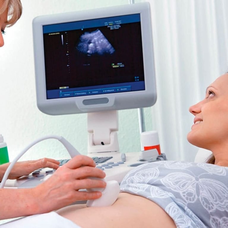
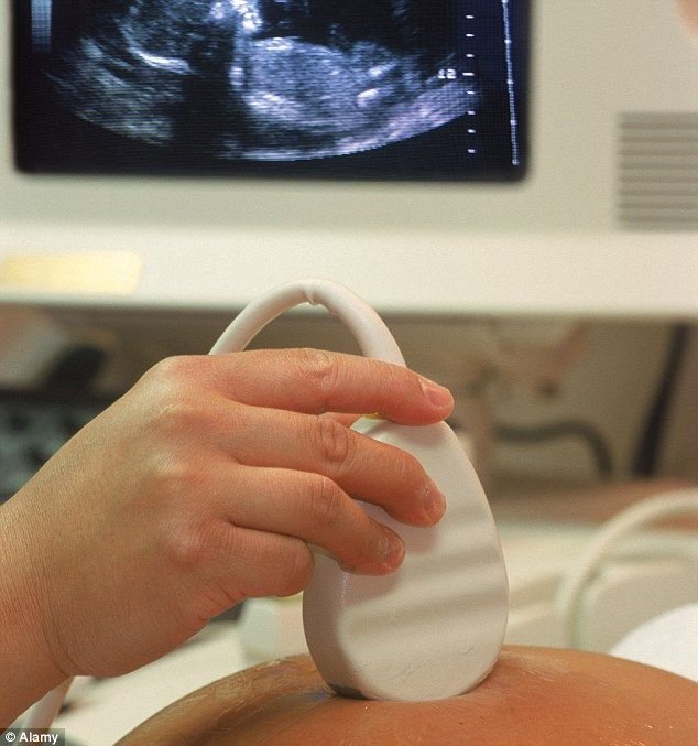
Early Pregnancy Scan
An Early Pregnancy Scan is an ultrasound performed during the first trimester, typically between 6 and 12 weeks of pregnancy. It helps confirm the pregnancy, assess the baby’s development, determine the due date, and identify any early concerns.
Benefits:
- Confirms the pregnancy is inside the uterus.
- Detects the baby’s heartbeat.
- Estimates the gestational age and expected due date.
- Identifies single or multiple pregnancies.
- Helps detect early pregnancy complications.
- Provides reassurance about your baby’s early development.
NT Scan (Nuchal Translucency Scan)
A Nuchal Translucency (NT) Scan is a specialized ultrasound performed during the 11 to 13 weeks + 6 days of pregnancy. It measures the fluid-filled space at the back of the baby’s neck to help assess the risk of certain chromosomal conditions and evaluates early fetal development.
Benefits:
- Screens for the risk of chromosomal abnormalities.
- Assesses early fetal growth and development.
- Helps detect certain structural abnormalities.
- Confirms gestational age and expected due date.
- Provides valuable information for further prenatal care.
- Safe, non-invasive, and radiation-free.
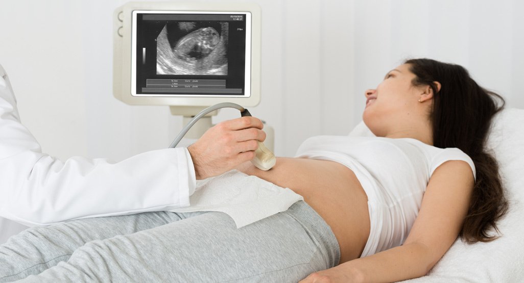

TIFFA Scan
A TIFFA Scan (Targeted Imaging for Fetal Anomalies) is a detailed ultrasound performed during the 18 to 22 weeks of pregnancy. It is also known as the Level 2 anomaly scan. This scan carefully examines the baby’s anatomy to check for structural development and identify any congenital abnormalities.
Benefits:
- Detailed evaluation of fetal organs and structures.
- Detects congenital anomalies at an early stage.
- Assesses growth of the baby in the second trimester.
- Checks placenta position and amniotic fluid level.
- Evaluates fetal heartbeat and movements.
- Helps in planning further pregnancy care if needed.
Doppler Scan
A Doppler Scan is a specialized ultrasound that evaluates blood flow in the baby, placenta, uterus, and umbilical cord. It helps doctors understand how well oxygen and nutrients are being supplied during pregnancy by measuring the speed and direction of blood flow in blood vessels.
Benefits
- Assesses blood flow to the baby and placenta.
- Helps detect pregnancy complications early.
- Monitors fetal well-being in high-risk pregnancies.
- Evaluates growth restriction in the baby (IUGR).
- Checks umbilical cord and uterine artery circulation.
- Guides timely medical decisions for mother and baby safety.
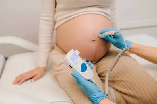

3D/4D Scan
A 3D/4D Scan is an advanced ultrasound that creates three-dimensional (3D) images and real-time moving images (4D) of the baby inside the womb. It provides a clearer view of the baby’s face, movements, and physical features compared to a standard ultrasound.
3D Scan: Still, detailed images of the baby
4D Scan: Live video-like movement of the baby in real time
Benefits:
- Gives a clear, detailed view of the baby’s face and body.
- Helps parents bond with the baby before birth.
- Assists doctors in evaluating fetal growth and anatomy.
- Detects certain external abnormalities.
- Monitors baby’s movements in real time.
- Provides reassurance about fetal development.
Advanced Ultrasound Technology
We use advanced ultrasound technology to deliver high-quality imaging for accurate diagnosis and comprehensive pregnancy monitoring. Our modern equipment provides clear, detailed images while ensuring a safe, comfortable, and non-invasive experience for every patient.
Key Features
- High-resolution imaging for exceptional clarity.
- Advanced 2D, 3D, and 4D ultrasound capabilities.
- Color and Spectral Doppler for blood flow assessment.
- Early and accurate detection of fetal and gynecological conditions.
- Safe, radiation-free imaging for mother and baby.
- Fast image processing and reliable diagnostic support.
Ultrasound Care Guide
Simple instructions for a smooth scan experience and accurate results.
- Carry Previous Reports
- Drink Water (if advised)
- Comfortable Clothing
- Follow Doctor Instructions
- Collect Reports
- Consult Doctor
- Schedule Next Scan
- Maintain Pregnancy Record
Dedicated Specialists For Your Care
Experienced doctors providing compassionate pregnancy care, advanced ultrasound services, and accurate diagnostic imaging.

FAQs
Your IVF questions, clearly answered here
Find clear, trusted answers to the most common questions about IVF—designed to guide and support you every step of the way.
Curious About IVF? Let's Talk.

Call Us:

E-mail Us:
Yes. Ultrasound uses sound waves instead of radiation, making it a safe and reliable method for monitoring your baby's growth and development throughout pregnancy.
Your first pregnancy scan is usually recommended between 5–8 weeks to confirm pregnancy, check the baby's heartbeat, and estimate your due date.
An NT Scan is performed between 11–14 weeks to measure the fluid behind your baby's neck and assess the risk of certain chromosomal abnormalities.
The NT Scan screens for chromosomal abnormalities in early pregnancy, while the TIFFA Scan is a detailed anatomy scan that evaluates your baby's organs and development.
A Doppler Scan is recommended when your doctor needs to evaluate blood flow between the placenta, umbilical cord, and baby, especially in high-risk pregnancies.
A TVS Scan is generally not painful. Some women may experience mild discomfort, but the procedure is safe, quick, and commonly used during early pregnancy.
A 3D/4D Scan is usually recommended between 24–32 weeks to capture detailed images of your baby's facial features and movements.
Most pregnancy ultrasound scans take between 15–30 minutes, depending on the type of scan and your baby's position.
Preparation depends on the scan type. Some ultrasound examinations require a full bladder, while others do not. Your doctor will provide instructions beforehand.